

Diagnostic Portable B/W MSK Ultrasound Systems
Diagnostic Portable B/W Musculoskeletal Ultrasound Scan
What are features of our B/W ultrasound machine?
Ergonomic Design
Up to 3 Transducer Ports
Light Weight and Compact
Large Anti-flickering HD LED Screen
Tilting Monitor Angle Adjustment
Backlit Keyboard and Intelligent Panel
Long-lasting Battery for 90 mins
Efficient Diagnosis
μ-Scan – Speckle reduction & Edge Enhancement
Spatial Compound Imaging
PIH – Pure Inversion Harmonic
Wide Scan – Image Area Enlargement
PW Doppler
Chroma – Highlight Image Details
B Steer for Linear Probe
Ease of Use
Quick Boot Up
Auto Brightness Adjustment
Auto Image Optimized
Auto IMT
Auto Trace
Expansive Accessories
Wi-Fi and Bluetooth Available
DICOM 3.0
500GB Hard Disk
Height Adjustable Trolley
Durable, Carry-on Site Suitcase
What is operation mode of our msk ultrasound systems?
| Operation Mode | B |
| Dual B | |
| Quad B | |
| Pure Inversion Harmonic Imaging ( PHI ) | |
| Trapezoid Imaging (Linear And Convex) | |
| Real-time Panoramic Imaging(B mode ) | |
| Panoramic support in Linear and convex. Velocity indicator and image rotate. | |
| M | |
| PW CW | |
| Duplex: B/M, B/PW, can be defined in new preset | |
| HD Zoom | |
| Compound Imaging (0,1,2,3,4) | |
| u-scan(5 levels adjustment) | |
| Needle enhancement ( Vis-needle ) |
| Cine Loop | available for all mode |
| Retrospective Storage | 27s retrospective saved in real time |
| Prospective Storage | 480s prospective saved in real time |
| Freeze Storage | required |
| Image Format | JPG, BMP, TIF |
| Clipboard function | is avilibale in frozen B mode |
| Clip Format | WMV, AVI |
| One Key Save | required |
| Doppler cine playback | speed is adjustable, sound can be played back |
| Print&Store Region | Full Screen, Image&Result Area, US Image |
| Image Share Service | required |
| Store To UsbDisk | required |
| Storage Capability | Hard Disk can up to 500G/1T; SSD can up to 128GB |
| Report Tables | Abdominal, OB/Gyn, Cardiology, Urology, Small Parts, Vascular,IMT |
| Report Format | TXT, PDF |
| Report Templet | at least 6 images can be displayed in report |
What are configuration of our portable MSK ultrasound?
| Operation Mode | B Dual B Quad B Pure Inversion Harmonic Imaging ( PHI ) Trapezoid Imaging (Linear And Convex) Real-time Panoramic Imaging(B mode ) Panoramic support in Linear and convex. Velocity indicator and image rotate. M PW CW Duplex: B/M, B/PW, can be defined in new preset HD Zoom Compound Imaging (0,1,2,3,4) u-scan(5 levels adjustment) Needle enhancement ( Vis-needle ) |
| Standard Hardware | E1 main unit 15.6"High Resolution LCD color monitor (with auto-adaptive LED backlight) One transducer connector USB 2.0/Hard Disk 500 G Adaptor |
| Standard Software | Imaging modes: B/ 2B/ 4B/ M Dynamic Multi-beam Technology Tissue Harmonic Imaging Pure Inversion Harmonic Imaging Tissue Specification Imaging Spatial Compound Imaging Widescan: Trapezoid Imaging μ-Scan: 2D speckle reduction technology TEI Index DICOM 3.0: Store, Send Auto: Auto optimization forB/ M/ PW TGC: Time gain compensation Zoom Standby Mode Show Galary 2D Steer Biopsy Guide |
| Stardard Configured Transducers | 96 element linear L746 (Vascular, Small Parts, MSKetc.),4.0-12.0MHz/ 50mm |
| Optional Transducers | 96 element convex C361 (Abdominal, Obstetrics, Gynecology) 2.0-6.0MHz/ R60mm 96 element linear L746 (Vascular, Small Parts, MSK etc.), 4.0-12.0MHz/ 50mm 128 element endocavity EC9-5 (Gynecology, Obstetrics, Urology), 3.0-15.0MHz/R8mm 128 element convex C351 (Abdominal, Obstetrics, Gynecology), 2.0-6.0MHz/ R50mm 128 element micro-convex array C613 (Cardiology, Pediatrics), 4.0-13.0MHz/ R14mm 128 element endocavity 6V1 (Gynecology, Obstetrics, Urology), 3.0-15.0MHz/ R11mm 128 element linear array L741(Vascular, Small Parts, MSK etc.), 4.0-16.0MHz/ 46mm |
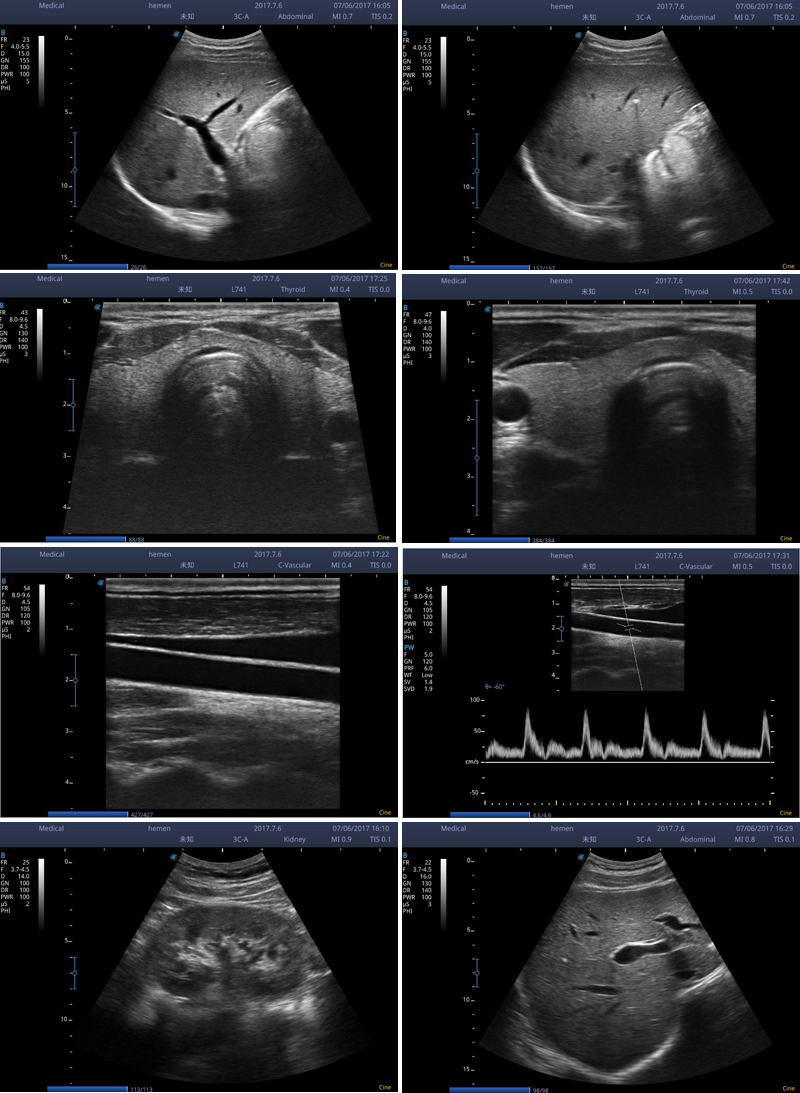
Demo images of our B/W MSK ultrasound scan