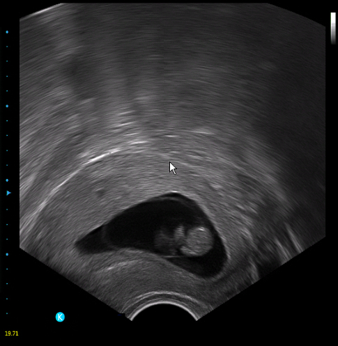


MCL-K0 Portable Color Doppler Ultrasound Scanner
MCL-K0 Portable Color Doppler Ultrasound Scanner
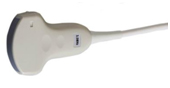
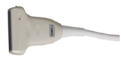
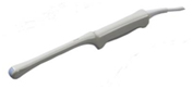
Features Probes 5 Steps Multi-frequency Image 3.5Mhz abdominal probe 2.0, 3.0, 3.5, 4.0, 5.5Mhz 7.5Mhz linear probe 6.0, 6.5, 7.5, 10.0, 12.0Mhz 6.5Mhz transvaginal probe 5.0, 6.0, 6.5, 7.5, 9.0Mhz B,B/B,4B,B/M,M, B/C,B/C/D,B/D, duplex, triplex, CFM, PW Signal processing: Full-digital beam forming, dynamic filter, dynamic real time receiving focusing, spectral processing, CFM processing, real-time dynamic focusing, dynamic aperture in all fields Image processing: THI Storage: 16G Power adjustable Smoothing function Edge enhancement One-key optimization Image conversion Wall filter adjustable Base line adjustable PRF adjustable AIO-Auto image optimization IZoom: Instant full screen image I-Image: intelligent optimization MBF: Multi Beam Former SA: Synthetic Aperture Ultrasonic imaging Iclear: Speckle Noise Reduction CDF: Contunuous Dynamic Focusing General measurement Normal, MSK, ABD, OB, Pelvic,Urology, Cardiac, Small Parts, Vascular Normal measurement Volume,V3L,STD_S,Area Trace,Mtime,MHR,D Time,DV,D Common,D Auto,Area,Angle,CrossLine,STD D,ParalleLine,Mdist,MV,D,DA,D Trace ABD packages ABD, Aorta, R_Kidney&L_Kidney, Bladder, Prostate Vascular Stenosis D, senosis A, Intima, Arterial, Venous MSK Distance, Area,Hip_Angle Scanning depth ≥250mm Probe elements 80 Cine loop Automatically & manually Image storage format BMP, JPEG, PNG,DICOM(Option) Input/output ports Video Port, S-Video Port, Remote Port, LAN1/2 Port, VGA Standard Configuration Main unit, 12 inch LED monitor, 3.5Mhz convex probe, 7.5Mhz linear probe, 2 Probe connectors, User’s Manual, hard disk (SSD) Options 6.5Mhz TransvaginalProbe, 3.5Mhz Micro-convex Probe, Trolley, Printers,Biopsy kit,Aluminum case
Smart design
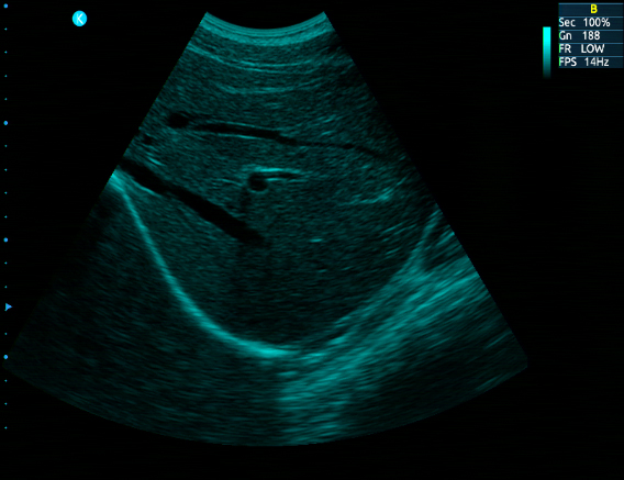
High Resolution Imaging System
Easy-operation Ergonomic Design
Better Optimize Image Quality
Smart and Light -Weight Design
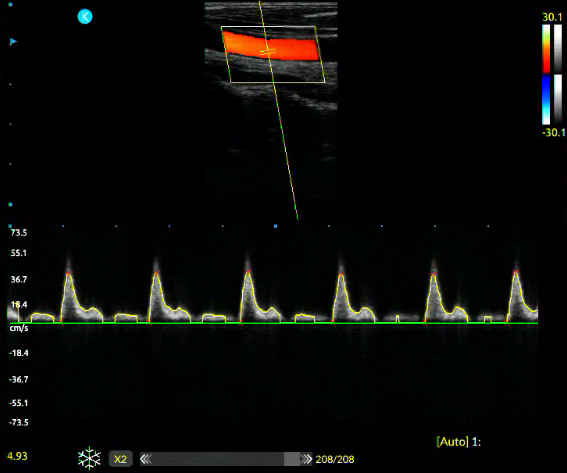
PW: Pulse Wave Doppler
The launch and reception of Ultrasonic pulse wave are processed by one probe, and start to receive echo signal after delay of schedule time.
Pseudo:
Pseudo color processing will change the gray level image to color image. Pseudo color makes the users distinguish the different organs issue. KO' s Pseudo color is more than 15 kinds of different color.
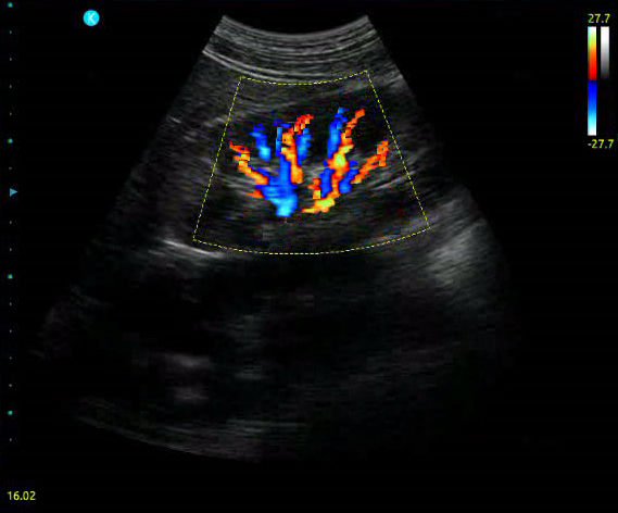
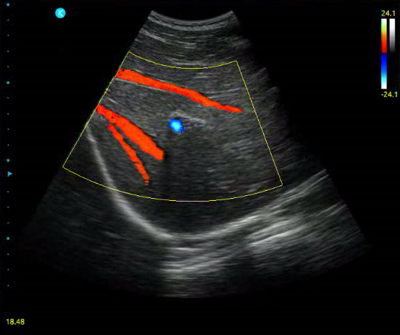
CF: Color Flow Mode
Double ultrasonic scanner system which could show B ode image and Doppler blood flow data(ie.blood flow irection, speed, velocity dispersion) simultaneously.
THI: Tissue Harmonic Imaging
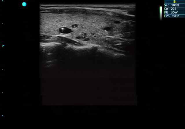
Tissue Harmonic imaging, which avoids many of the near-field artifacts that affect fundamental imaging. And have the advantage of good signal-noise ratio, better spatial resolution, enhance tissue contrast, Improve deep tissue echo information.
Probes information
Technical specifications